Search / Thermo Fisher Scientific
MA5-12557
p53 Monoclonal Antibody (DO-7) - MA5-12557
3 flagged images · first reported 17 May 2026 · by Anonymous; Sholto David; Sholto David (via Marvin Bader)
What was found
- Several bands are identical after rotation and flipping
- Immunofluorescence images are almost the same as image 119 despite representing a different clone (DO-7)
- Atypical background texture on the image using antibody from a competitor. Two antibodies were tested by Western blot. The left image shows the Thermo Fisher antibody (MA5‑12557, clone DO‑7) and the right image shows a competitor antibody (clone 1C12). The image on the right exhibits an unusually uniform background that lacks the pixel grain or background noise typically seen in western blot images.
The images
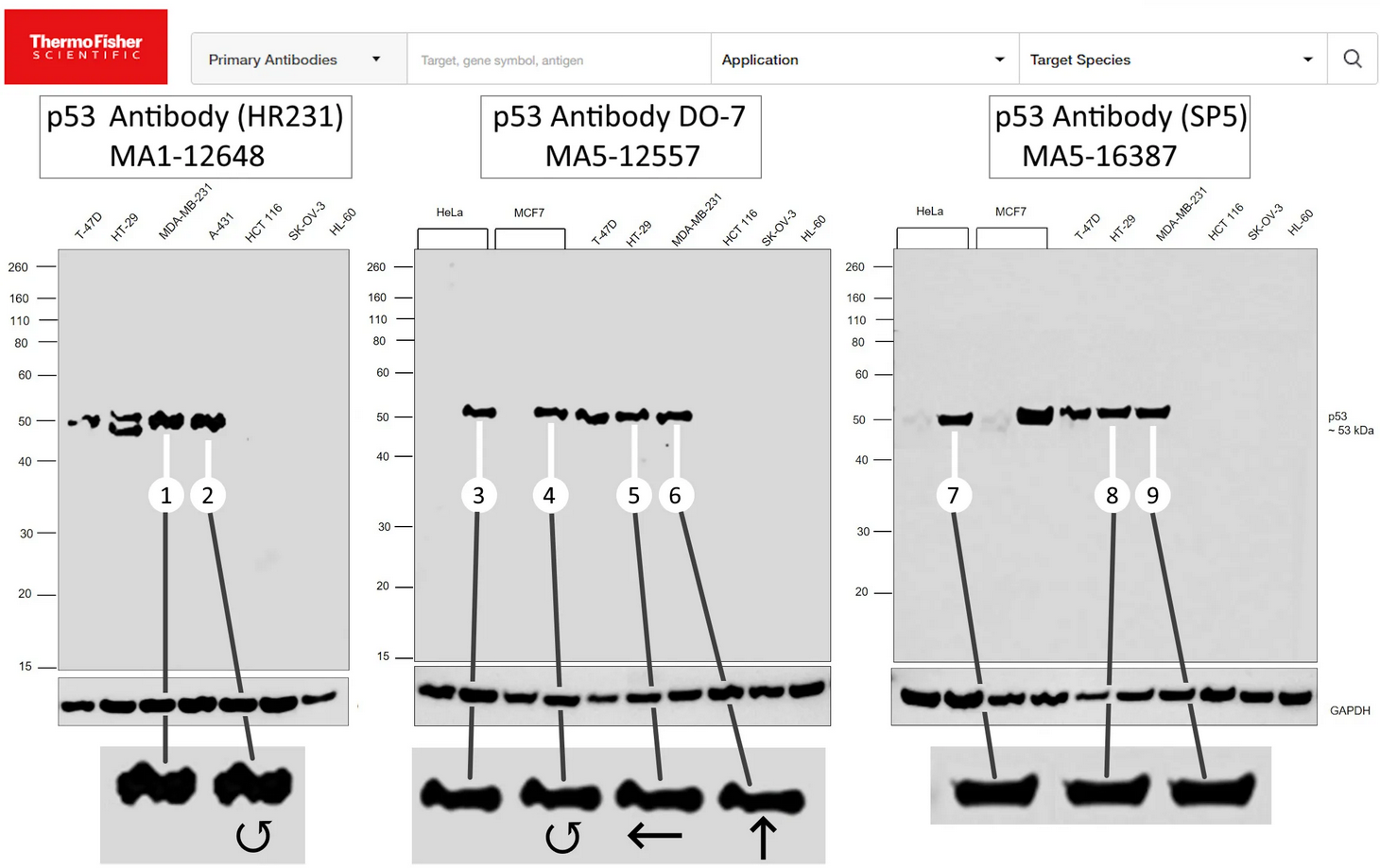
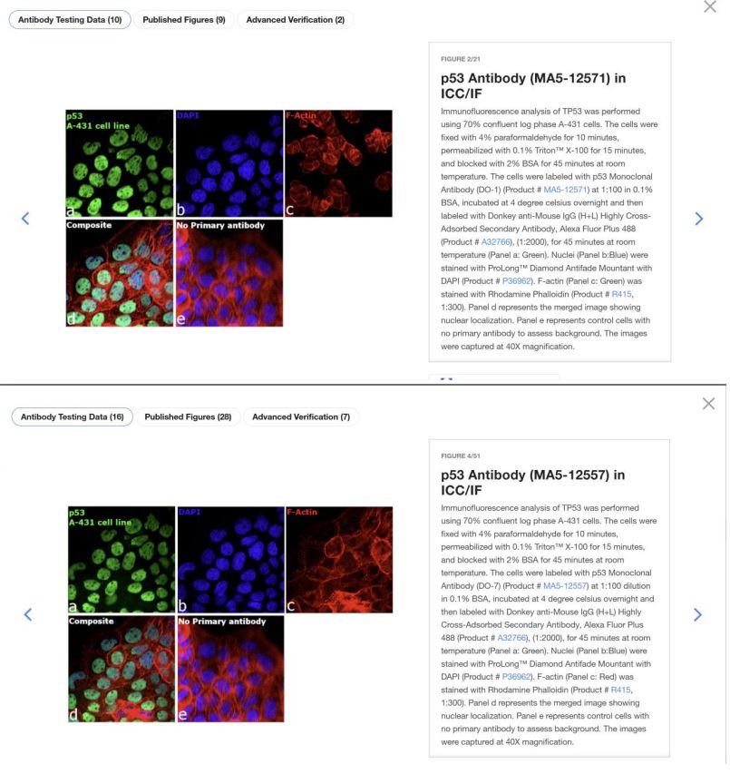
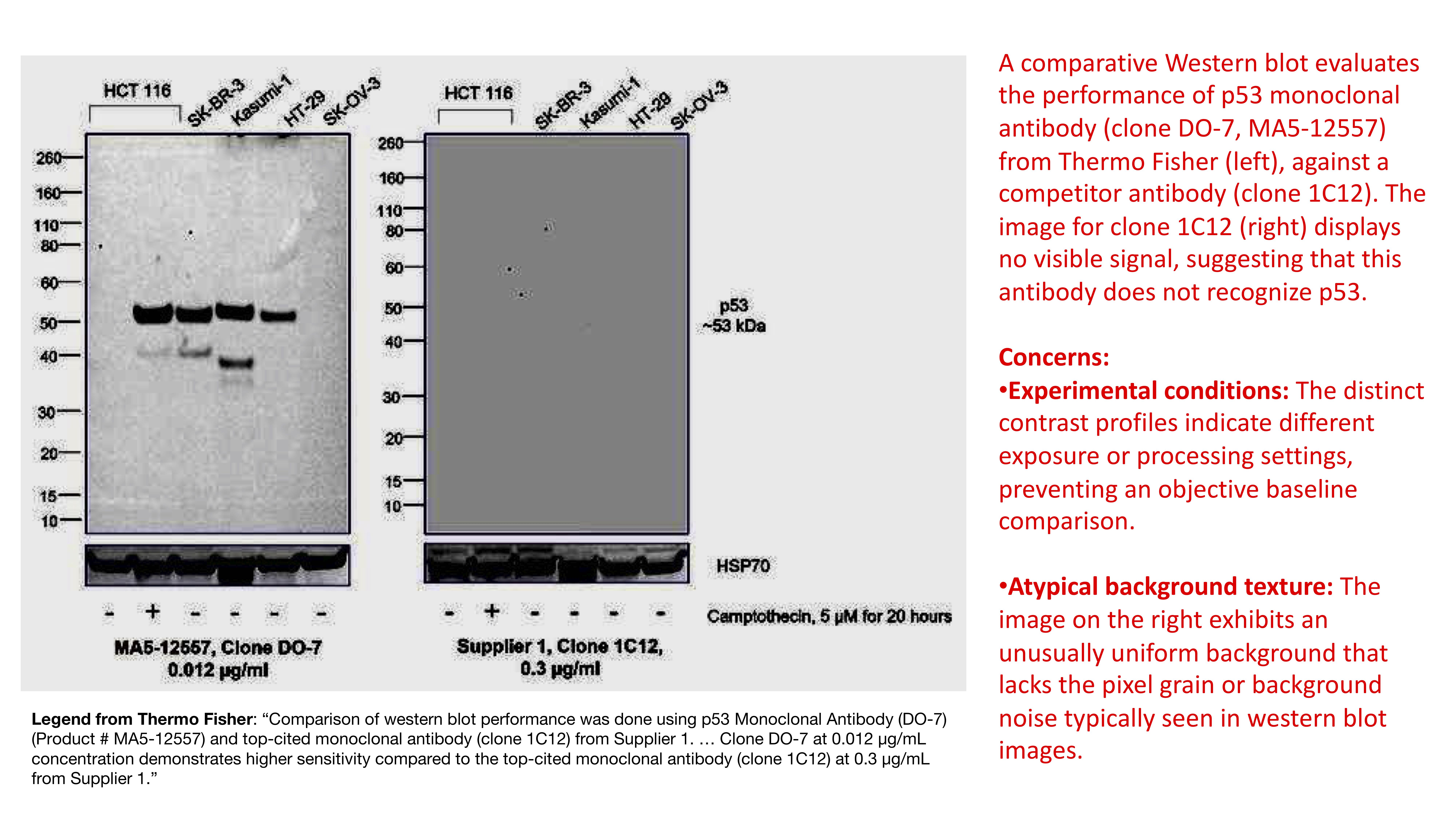
Annotations by the contributors listed above, republished under CC BY 4.0 from the Zenodo repository.
A flag on this page means the images the vendor published as evidence cannot be trusted as evidence. Unless an independent knockout test is shown above, it says nothing about whether this antibody binds its target. Treat it as untested until you or someone else controls it.